Our group

Human pluripotent stem cell differentiated retinal ganglion cells (hRGCs)

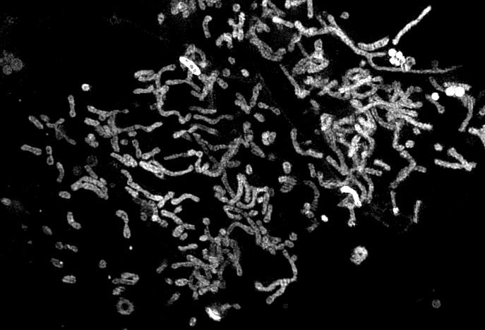
Structural illumination microscopy (SIM) image of mitochondria in hRGCs

Immunohistochemistry confocal image of mouse optic nerve head stained against mitochondria (Green), phospho-PGC1-alpha (Red) and nucleus (Blue)

Human embryonic stem cells at different cell-cycle stages revealed by immunofluorescence against Ki-67 protein (magenta), actin cortex (green)